 Loading... Please wait...
- Call us on 443-686-9618
- Wish Lists
- My Account
- 0.00
Categories
- Home
- IHC Antibodies
- APAF1 IHC Antibody
APAF1 IHC Antibody
Product Description
Description |
Apoptotic peptidase activating factor 1, also known as APAF1, is a protein which in humans is encoded by the APAF1 gene. This gene is mapped to chromosome 12q23. It encodes a cytoplasmic protein that initiates apoptosis. And it is an essential downstream effector of p53-mediated apoptosis.This protein contains several copies of the WD40 repeat domain, a caspase recruitment domain (CARD), and an ATPase domain (NB-ARC). In the presence of cytochrome c and dATP, APAF1 assembles into an oligomeric apoptosome, which is responsible for activation of procaspase-9 and maintenance of the enzymatic activity of processed caspase-9. Furthermore, APAF1 is inactivated in metastatic melanomas, leading to defects in the execution of apoptotic cell death. Additionally, APAF1 has been shown to interact with NLRP1,5 Caspase-9, APIP, BCL2-like 1, and HSPA4. |
Catalog Number |
IW-PA1249 |
Quantity |
9 ml |
|
Host |
Rabbit |
|
Clone |
Polyclonal |
|
Isotype |
Rabbit IgG |
|
Immunogen |
A synthetic peptide corresponding to a sequence at the N-terminal of human APAF1, identical to the related rat and mouse sequence. |
|
Purity |
Immunogen affinity purified. |
|
Conjugate |
Unconjugated |
|
Species Reactivity |
Human, mouse, rat. Not tested in other species. |
|
Positive Control |
Rat brain |
|
Cellular Localization |
Cytoplasmic |
|
Form |
Ready to use solution in PBS with stabilizer and 0.01% sodium azide. No further dilution needed. Serum blocking step should be omitted. |
|
Storage |
Store at 2-8 °C. Do not freeze. |
|
Applications |
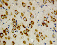
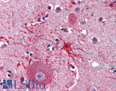
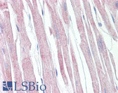
IHC-P: Heat induced epitope retrieval (HIER) is required on formalin fixed paraffin embedded sections. IHC-Fr: Not tested. ICC: Not tested. |
|
Limitations |
This product is intended for Research Use Only. Interpretation of the test results is solely the responsibility of the user. |
|
Precautions |
Users should follow general laboratory precautions when handling this product. Wear personal protective equipment to avoid contact with skin and eyes. |
|
References |
1. Kim, H.; Jung, Y. K.; Kwon, Y. K.; Park, S. H. : Assignment of apoptotic protease activating factor-1 gene (APAF1) to human chromosome band 12q23 by fluorescence in situ hybridization. Cytogenet. Cell Genet. 87: 252-253, 1999. 2. Robles, A. I.; Bemmels, N. A.; Foraker, A. B.; Harris, C. C. : APAF-1 is a transcriptional target of p53 in DNA damage-induced apoptosis. Cancer Res. 61: 6660-6664, 2001. 3. Bao, Q.; Lu, W.; Rabinowitz, J. D.; Shi, Y. : Calcium blocks formation of apoptosome by preventing nucleotide exchange in Apaf-1. Molec. Cell 25: 181-192, 2007. 4. Soengas, M. S.; Capodieci, P.; Polsky, D.; Mora, J.; Esteller, M.; Opitz-Araya, X.; McCombie, R.; Herman, J. G.; Gerald, W. L.; Lazebnik, Y. A.; Cordon-Cardo, C.; Lowe, S. W. : Inactivation of the apoptosis effector Apaf-1 in malignant melanoma. Nature 409: 207-211, 2001. 5. Chu, Z L; Pio F, Xie Z, Welsh K, Krajewska M, Krajewski S, Godzik A, Reed J C (Mar. 2001). "A novel enhancer of the Apaf1 apoptosome involved in cytochrome c-dependent caspase activation and apoptosis". J. Biol. Chem. (United States) 276 (12): 9239-45. 6. Cho, Dong-Hyung; Hong Yeon-Mi, Lee Ho-June, Woo Ha-Na, Pyo Jong-Ok, Mak Tak W, Jung Yong-Keun (Sep. 2004). "Induced inhibition of ischemic/hypoxic injury by APIP, a novel Apaf-1-interacting protein". J. Biol. Chem. (United States) 279 (38): 39942-50. 7. Li, P; Nijhawan D, Budihardjo I, Srinivasula S M, Ahmad M, Alnemri E S, Wang X (Nov. 1997). "Cytochrome c and dATP-dependent formation of Apaf-1/caspase-9 complex initiates an apoptotic protease cascade". Cell (UNITED STATES) 91 (4): 479-89. 8. Hu, Y; Benedict M A, Wu D, Inohara N, Núñez G (Apr. 1998). "Bcl-XL interacts with Apaf-1 and inhibits Apaf-1-dependent caspase-9 activation". Proc. Natl. Acad. Sci. U.S.A. (UNITED STATES) 95 (8): 4386-91. 9. Pan, G; O'Rourke K, Dixit V M (Mar. 1998). "Caspase-9, Bcl-XL, and Apaf-1 form a ternary complex". J. Biol. Chem. (UNITED STATES) 273 (10): 5841-5. 10. Saleh, A; Srinivasula S M, Balkir L, Robbins P D, Alnemri E S (Aug. 2000). "Negative regulation of the Apaf-1 apoptosome by Hsp70". Nat. Cell Biol. (ENGLAND) 2 (8): 476-83.
|

