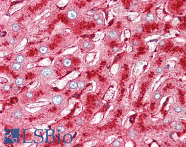
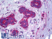
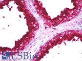
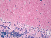
Specificity: Used for diagnostic by staining of lysosomes/melanosomes. Directed against a predominantly intracellular protein of human melanoma cells. The antigen is abundantly present in malignant melanomas, nevocellular nevi, and neuroendocrine tumors, whereas it has not been detectable in most other tissues. The antigen recognized is a glycoprotein which appears as a broad band in SDS-PAGE. The antigen was shown to consist of a single protein backbone to which two or three V-linked glycans were added cotranslationally. Extensive further heterogeneity was generated in the Colgi compartmen and was shown to be dependent on the presence of complex type sugars. Although the antigen is associated with melanomas, it was not codistributed with the tyrosinase activity associated with melanogenesis. The antigen did show codistribution with cathepsin D, which is a marker for lysosomal functions.This antibody was originally described as a marker for melanoma. Recently, it resurfaced as a marker to separate cellular neurothekeoma from other dermal tumors in the differential diagnosis.